Back Of Skull Anatomy - Base of Skull from Above | ClipArt ETC : William is a final year medical student in australia who has taught anatomy to tertiary science and.
Back Of Skull Anatomy - Base of Skull from Above | ClipArt ETC : William is a final year medical student in australia who has taught anatomy to tertiary science and.. The skull is the bony skeleton of the head. This anatomic region is complex and poses surgical challenges for otolaryngologists and neurosurgeons alike. From an anatomical perspective, the skull is divided into two parts: Please feel free to download and print. The bbc is not responsible for the content of external websites.
Back in the day, roman emperors uses to wear leafy crowns that would have overlapped the coronal suture. The skull is the bony skeleton of the head. The frontal, parietal, temporal and occipital bones are joined at the cranial sutures. .back of skull, bone spur back of skull, skull bone back of head, bone, bone back of head bigger on one side, bone back of the head, bone on back human anatomy bones human anatomy bone kit, human anatomy bones games, human anatomy coccyx bone, human anatomy collarbone, learn. A cartilaginous mould begins to grow this is why raising your eyebrows can create the appearance that the back of the head is moving.
Anatomy of the Skull Base and Related Structures: Elements ... from neupsykey.com Anatomy of the skull and bones of cranium on medical illustrations. The skull is a bony structure that supports the face and forms a protective cavity for the brain. The base of the skull (or skull base) forms the floor of the cranial cavity and separates the brain from the structures of the neck and face. This is a model of the human (homo sapiens) skull. It is comprised of many bones, formed by intramembranous ossification, which are joined together by sutures (fibrous joints). Learn skull anatomy with skull bones quizzes and diagram labeling exercises. The skull is the bony skeleton of the head. The skull or known as the cranium in the medical world is a bone structure of the head.
The skull has a single occipital condyle.7 the skull consists of five major bones:
The skull begins to form prior to week 12 of embryogenesis. Anatomical structures of the skull include: The base of the skull (or skull base) forms the floor of the cranial cavity and separates the brain from the structures of the neck and face. Skull bones aren't fused together at birth. In order to be light, the skull is made up by flat and irregular bones, and has hollow spaces called the sinuses. They don't move and united into a single unit. It was then cleaned, adapted and polypainted this model is part of a comparison with the skull of a human. Anatomy of the skull and bones of cranium on medical illustrations. The bbc is not responsible for the content of external websites. A thorough description is beyond the. Axial muscles of the head, neck, and back. These joints fuse together in adulthood. The skull includes the upper jaw and the cranium.
Understanding the human skull anatomy is necessary for a wide range of professionals from doctors (dentists, oral surgeons, neurosurgeons, etc.) to the structure of the skull bones is to a large extent determined by and interconnected with the anatomy of the sensory organs, situated in the head, as. Anatomy next provides anatomy learning tools for students and teachers. It supports and protects the face and the brain. Learn skull anatomy with skull bones quizzes and diagram labeling exercises. The skull begins to form prior to week 12 of embryogenesis.
Free photo: Skull - Anatomy, Human, Spooky - Free Download ... from jooinn.com So, the human skull consists of 23 bones. Learn about skull base anatomy with free interactive flashcards. This anatomic region is complex and poses surgical challenges for otolaryngologists and neurosurgeons alike. The skull or known as the cranium in the medical world is a bone structure of the head. The skull includes the upper jaw and the cranium. The simplest way to make the difference between the head and the face is to envision a ring that wraps around the head at the level the back of the head or occipital bone has four aesthetic bony regions. Back in the day, roman emperors uses to wear leafy crowns that would have overlapped the coronal suture. Human skull from the front.
Excluding ear ossicles, it is made of 22 bones.
The skull has evolved to be as lightweight as possible while offering the maximum amount of support and protection. Learn more about the anatomy and function of the skull in humans and other vertebrates. Skull bones aren't fused together at birth. Looking at it from the inside it can be subdivided into. Skull reshaping is done on any of the structures that lie above the face. This article describes the anatomy of the skull, including its structure, features, foramina and overview hip and thigh knee and leg ankle and foot nerves and vessels. The skull is the bony skeleton of the head. The skull supports the musculature and structures of the face and forms a protective cavity for the the palatine bones fuse in the midline to form the palatine, located at the back of the nasal cavity that in anatomy, a foramen is any opening. In order to be light, the skull is made up by flat and irregular bones, and has hollow spaces called the sinuses. This anatomic region is complex and poses surgical challenges for otolaryngologists and neurosurgeons alike. Human skull from the front. The major sutures are the coronal suture, sagittal suture, lambdoid suture and squamosal sutures. The skull performs vital functions.
The skull has a single occipital condyle.7 the skull consists of five major bones: The skull or known as the cranium in the medical world is a bone structure of the head. The skull bones can be classified into two groups: Excluding ear ossicles, it is made of 22 bones. Skull, skeletal framework of the head of vertebrates, composed of bones or cartilage, which form a unit that protects the brain and some sense organs.
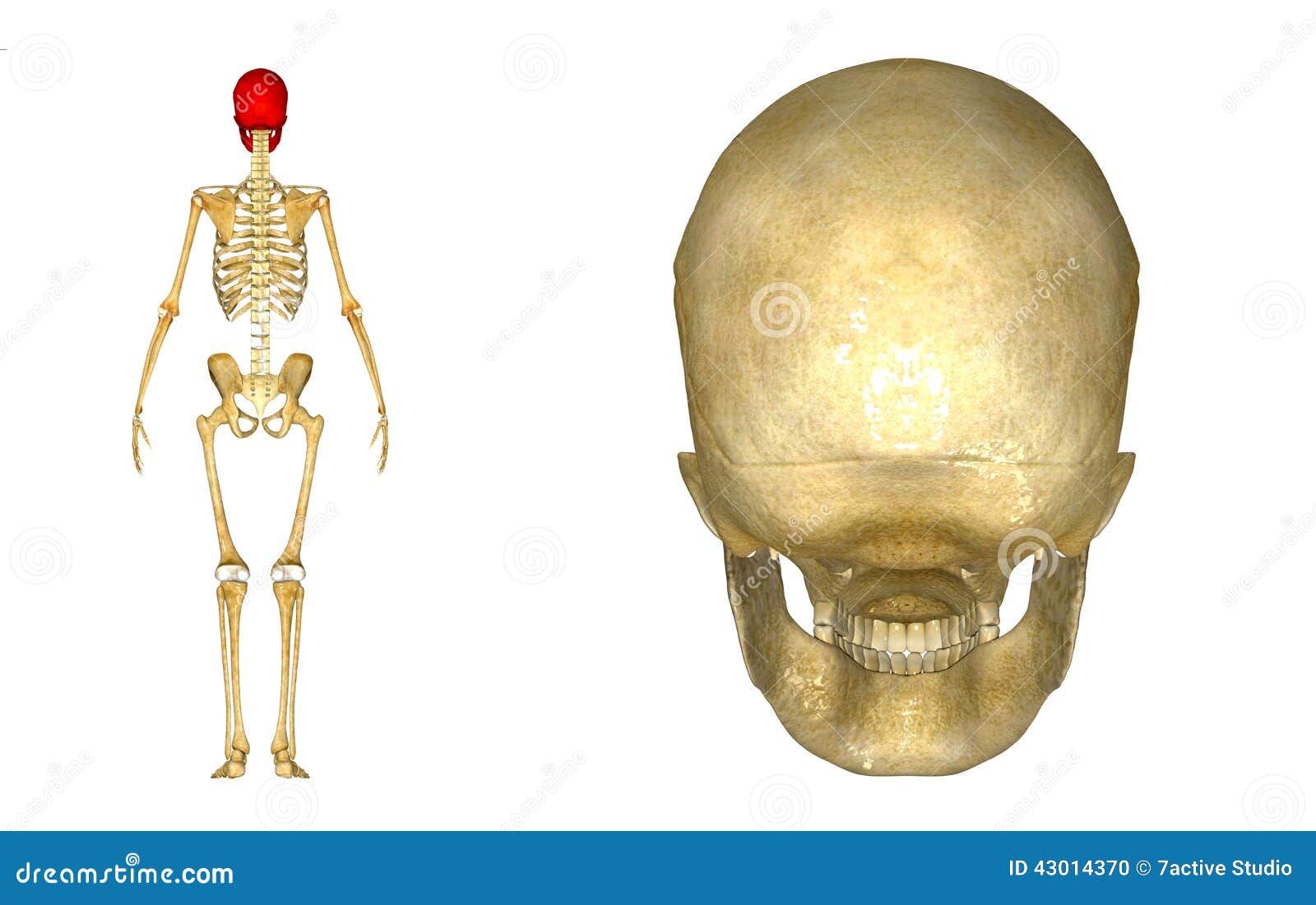
Human Skull Back Stock Illustration - Image: 43014370 from thumbs.dreamstime.com Learn more about the anatomy and function of the skull in humans and other vertebrates. It is comprised of many bones, formed by intramembranous ossification, which are joined together by sutures (fibrous joints). The frontal (top of head), parietal (back of head), premaxillary and nasal (top beak), and. It supports and protects the face and the brain. The skull performs vital functions. The skull is a bony structure that supports the face and forms a protective cavity for the brain. This is a model of the human (homo sapiens) skull. Anatomy of the skull and bones of cranium on medical illustrations.
It was then cleaned, adapted and polypainted this model is part of a comparison with the skull of a human.
From an anatomical perspective, the skull is divided into two parts: The base of the skull (or skull base) forms the floor of the cranial cavity and separates the brain from the structures of the neck and face. Inferior view of base of the skull. A cartilaginous mould begins to grow this is why raising your eyebrows can create the appearance that the back of the head is moving. Anatomy of the skull and bones of cranium on medical illustrations. Learn about the anatomy of the skull bones and sutures as seen on ct images of the brain. The skull is the bony skeleton of the head. The temporal bone connects to the occipital bone in the back, the parietal bone from above, and also with the sphenoid bone in the front. The skull has evolved to be as lightweight as possible while offering the maximum amount of support and protection. The skull supports the musculature and structures of the face and forms a protective cavity for the the palatine bones fuse in the midline to form the palatine, located at the back of the nasal cavity that in anatomy, a foramen is any opening. Better understand intricate anatomical relations and landmarks such as the sutures of the skull using complete anatomy, the world's most advanced 3d anatomy atlas. The skull begins to form prior to week 12 of embryogenesis. The skull has a single occipital condyle.7 the skull consists of five major bones:


0 Komentar